Connect the right patient to the right treatment, resources, and community.

Device Aided Therapies for Parkinson's Disease

  • Device-aided therapies for Parkinson’s disease (Subcutaneous Infusion, DBS, Focused Ultrasound, and Duopa) can be an effective option for many symptoms of Parkinson’s disease.
  • These options may be explored when a patient has tried numerous oral medications for PD without success, despite trying multiple medication adjustments with their neurologist or MDS.
  • When a patient’s tremor, motor fluctuations (on/off), and/or dyskinesia persist and significantly impact their quality of life, it may be time to consider device-assisted therapies. 

Indications for Device Aided Therapies

Subcutaneous Infusions

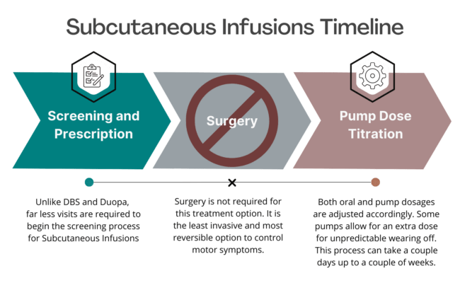
Introducing the potential for an upcoming Subcutaneous Infusion Pump designed to provide a steady dose of apomorphine directly under the skin to reduce OFF episodes and troublesome dyskinesia in Parkinson’s disease. Unlike other devices, this option is minimally invasive, discreet, and easily concealed, and does NOT require surgery or hospitalization.

Patients have the flexibility to remove the pump whenever they choose (i.e. in the shower) offering a level of convenience and control over their treatment regimen. This therapy is both easy to start and stop, allowing patients to explore its benefits without the commitment of irreversible surgery, making it a significant alternative to considering Deep Brain Stimulation (DBS). 

Clinical studies determined that this Infusion Pump reduced OFF time by an average of 3 hours with a similar increase in good ON time without troublesome dyskinesia. Once approval is gained, this pump will be the first and only continuous infusion device available for PD. 

  • Subcutaneous therapy allows for a steady flow of medication throughout the day. The continuous rate settings will provide most of the daily medication, and you may give yourself extra doses with the pump if needed. Generally, symptoms do not improve beyond what a patient considers good “On” time before subcutaneous therapy.

  • Some people choose to continue their other Parkinson’s medications (such as dopamine agonists or amantadine), although some patients may be able to stop these medications.
  • Patients may experience pain at the insertion site, particularly when changing the needle. Pain tends to be mild and resolves quickly. The main more serious side effect is an infection at the infusion pump site. Some patients may develop very small painless bumps (nodules) underneath the skin in previous infusion sites as well.

  • The side effects of treatment with subcutaneous medication are the same as those for the sublingual and intermittent subcutaneous injection form and include nausea, subcutaneous nodules orthostatic hypotension, dyskinesia, sleepiness, confusion, and hallucinations.

Subcutaneous infusion therapy is not a cure for Parkinson’s Disease and it does not slow PD progression. It generally does not help any Parkinson’s disease symptoms that do not improve with your best medication “on” time.

  • Patients with advanced Parkinson’s disease who respond well to oral carbidopa/levodopa, but experience significant motor fluctuations for more than 3 hours of OFF time, despite adding another class of medications such as MAO-B inhibitors or dopamine agonists.

  • Patients who have to take their medications very frequently and have difficulty maintaining a regular dosing schedule.

  • Patients who are hesitant to undergo surgical device-aided therapies, either due to concerns about procedural risks or lack of reversibility. 

DBS

  • DBS requires (2) surgeries that are not required for subcutaneous infusions.
  • DBS requires minimal continual care from a patient after surgery. Subcutaneous infusion sites need to be changed regularly by the patient.
  • DBS is somewhat reversible in that stimulation can easily be turned off. Removing the hardware from the body would require surgery. Subcutaneous is easily reversible in that the catheter can be immediately removed from the skin.

Focused ultrasound

  • Focused ultrasound is a major medical procedure involving brain surgery. Subcutaneous levodopa is less invasive, as it just requires the insertion of a small needle under the skin.
  • Subcutaneous infusion requires adjustment with a neurologist to set the continuous dose, extra dose, and morning dose. These settings can be changed if necessary, even years later. Focused ultrasound is a single procedure, effects are immediate and cannot be adjusted.

Duopa

  • Duopa requires a medical procedure to insert the PEG-J tube. Subcutaneous levodopa is less invasive, as it just requires the insertion of a small needle under the skin.
  • Both Duopa and subcutaneous levodopa infusion require adjustment with a neurologist to set the continuous dose, extra dose, and morning dose. These settings can be changed if necessary, even years later.
  • Both therapies rely on the medication levodopa for their effect.
  • Both procedures are completely reversible. The stoma required in Duopa may take slightly longer to heal when compared to removing a needle for subcutaneous therapy.

Deep Brain Stimulation (DBS)

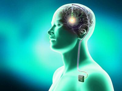
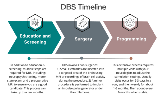
Deep brain stimulation (DBS) is a treatment for neurological conditions such as Parkinson’s disease, essential tremor, and epilepsy. DBS was first approved for the treatment of PD in 1997 and involves the surgical implantation of an electrode in the brain in an area important for the control of movement, specifically in a region called the globus pallidus internus (GPi) or the subthalamic nucleus (STN). Wires passing through the neck connect the electrode to a stimulator in the chest. The stimulator is a small pacemaker-like device that delivers a controlled electronic pulse to the brain.

Two regions in the brain may be chosen for electrode placement: the subthalamic nucleus (STN) or the globus pallidus internus (GPI). The area chosen depends on a patient’s symptoms. Both areas are very effective for a patient’s tremor and slowness. A clinician may be more likely to choose the STN if a patient experiences many adverse effects from carbidopa/levodopa such as excess dyskinesia. Stimulation of the STN often allows for greater medication reduction than stimulation of the GPi. There is some data to suggest that stimulation of the GPi leads to fewer problems with language and cognition. Many ongoing studies will help refine target choices for individual patients.

  • DBS is effective in treating the symptoms of Parkinson’s Disease such as slowness of movement, tremor, dystonia, or other symptoms that respond well to carbidopa/levodopa without causing excess involuntary movements, or dyskinesia.
  • Before ensuring a patient is appropriate for DBS, a clinician usually performs a “Levodopa Challenge Test” with a patient. During the test, the patient comes to the clinic without taking any medications. Over a few hours, the patient is given carbidopa/levodopa and repeatedly examined to evaluate the effect of the medications. Generally, the effect seen in the levodopa challenge test is a good indicator of the effect to expect from DBS. The one exception to this rule is medication refractory tremor. Often DBS is more effective for tremor than carbidopa/levodopa.
  • DBS stimulation is continuous, so it is very effective at treating motor fluctuations or the roller coaster of “on” and “off” symptoms many patients struggle with.
  • Risks of surgical procedure or hardware malfunction: The main risk of surgery is hemorrhage, or bleeding in the brain, which is a type of stroke. This risk is 1 to 2% for each side of the brain operated on. Infection may occur in up to 5% of patients at some time after surgery but most commonly in the first three months post-operatively. Generally, the infection involves the scalp or skin and infrequently involves the brain. Usually, patients can be treated with antibiotics, and sometimes hardware must also be removed temporarily, but rarely does the electrode in the brain need to be removed due to infection. The DBS device may stop working due to breaks in the wiring that connects the electrode to the pacemaker-like device in the chest. This risk is less than 5% in one’s lifetime. This would require additional surgery to replace or fix the DBS hardware.
  • Problems with speech: Some patients have reduced clarity of speech after DBS
  • Cognitive effects: A small number of people with PD have experienced a decline in memory and thinking after DBS surgery. This is why DBS is not appropriate for those patients with dementia.
DBS is not a cure for Parkinson’s Disease and it does not slow PD progression. It is generally not helpful for certain symptoms of PD such as problems with swallowing, speech, memory and thinking, balance, or freezing of gait.
  • Patients with Parkinson’s Disease for at least five years. DBS is not effective for disorders such as MSA, PSP, or CBD which mimic PD early in the disease.
  • Patients with “on/off” fluctuations despite consistent and regular medication dosing and despite trying additional medications to improve this.
  • Patients with troublesome dyskinesia despite medication adjustments.
  • Patients with troublesome dystonia despite medication adjustments.
  • Patients unable to tolerate carbidopa/levodopa due to side effects.
  • Patients with significant tremor that is not well controlled with medication, even despite medical management by a movement disorders specialist.
  • Patients without significant problems with memory and thinking that affect day-to-day life (dementia)

Focused Ultrasound:

  • DBS electrodes do not destroy healthy brain tissue. Instead, they create a temporary electrical signal that disrupts tissue activity. Focused ultrasound destroys brain tissue, creating a lesion in the brain.
  • DBS electrical stimulation can be adjusted over time as the symptoms of PD progress. Focused ultrasound cannot be adjusted after the initial procedure.
  • DBS requires significant programming after surgery and a time commitment for a patient and clinician. Focused ultrasound is a single procedure.
  • DBS requires a major brain surgery and a minor surgery. Both surgeries require anesthesia. Focused ultrasound requires one major brain surgery. The anesthesia required is minimal.
  • DBS is approved for both sides of the brain to improve symptoms on both sides of the body. Focused ultrasound is only approved for one side of the brain, to improve symptoms on one side of the body.

Duopa intestinal gel:

  • DBS uses electrical stimulation in the brain to improve PD symptoms. Duopa uses a continuous infusion of levodopa to improve these symptoms.
  • DBS requires a major brain surgery and a minor surgery. Duopa requires a minor stomach surgery to implant the tube.
  • Both DBS and Duopa can be adjusted over time to improve PD symptoms as the disease progresses.
  • DBS has a potential risk of worsening cognition. This is not true of Duopa.
    Subcutaneous levodopa:
  • DBS requires surgeries that are not required for subcutaneous levodopa infusions.
  • DBS requires minimal continual care from a patient after surgery. Subcutaneous levodopa infusion sites need to be changed regularly by the patient.
  • DBS is somewhat reversible in that stimulation can easily be turned off. Removing the hardware from the body would require surgery. Subcutaneous levodopa is easily reversible in that the catheter can be immediately removed from the skin.

Focused Ultrasound

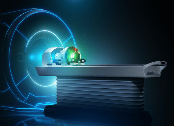
Focused Ultrasound therapy has emerged as a promising non-invasive treatment option for Parkinson’s disease, offering hope to patients grappling with its debilitating symptoms. By utilizing precisely targeted ultrasound waves to target areas of the brain, this approach aims to alleviate motor symptoms such as tremors, rigidity, and bradykinesia.

Unlike traditional surgical interventions, Focused Ultrasound therapy doesn’t involve incisions or anesthesia, minimizing the risk of complications and reducing recovery time. With ongoing research and clinical trials showing promising results in symptom management and improving quality of life, Focused Ultrasound therapy presents a potential breakthrough in the treatment landscape for Parkinson’s disease.

Preparation:

  • Your head will be shaved on the morning of the procedure to allow good contact between your head and the ultrasound.
  • You will be given some medicine through an IV to keep you comfortable during the procedure.
  • Your head will be placed in a head frame to keep your head steady and prevent it from moving. Next, a silicone membrane will be placed on top of your head. The membrane seals the space where cold water will circulate between your scalp and the helmet of the device. The water barrier helps keep your scalp cool and makes sure there is adequate contact between your head and the ultrasound equipment.
  • You will lie on an MRI bed, which slides in and out of the scanning area of the machine. The head frame is locked into position on the bed so you cannot move your head. Next, several scans will be taken of your brain. These scans help your physicians identify the area to be treated and the specific target area to aim the ultrasound waves.
  • Before treatment begins, you may be asked to draw spirals or write your name or perform certain hand and finger movements so your medical team can assess your tremor. You will be asked to repeat these tasks during various stages of treatment to see if the ultrasound is relieving your tremors.

Treatment:

  • Treatment begins with several short ultrasound pulses aimed at the target area. These low-energy (non-treatment level) pulses are administered to confirm that the proper target has been located.
  • Once the accuracy of the target is confirmed by MR images, the ultrasound energy is gradually increased over a series of stages. At each stage, the temperature of the targeted tissue is checked and MR images confirm that the procedure is continuing as planned. The MR images provide real-time feedback so the surgeon can make any needed adjustments.
  • You will be asked how you are feeling and will repeat hand and finger tasks to check treatment progress. Once your tremor is improving the ultrasound energy is increased so a small lesion forms.
  • You will be awake during the entire procedure inside the MRI and will be able to speak to your medical team. You will be given an emergency stop button to hold during the procedure. If you are experiencing pain or other adverse effects, you can push the stop button at any time.

Afterward:

  • The entire procedure, from preparation to getting off the table, takes about 3 to 4 hours.
  • After the procedure, you will move to a recovery room for observation. You may go home the same day or remain in the hospital for 24 to 48 hours. Your doctor will let you know when you can leave and when you need to return for a follow-up visit.
Focused ultrasound can be helpful for some of the symptoms of Parkinson’s Disease, predominantly tremor. The procedure is only approved for one side of the brain at present, so tremor would only improve on one side of the body.
Focused ultrasound is not a cure for Parkinson’s Disease and it does not slow PD progression. The procedure is not adjustable. It is generally not helpful for certain symptoms of PD such as problems with swallowing, speech, memory, and thinking, balance, or freezing of gait.
  • Temporary side effects of the procedure: The most common temporary side effects after the Focused Ultrasound procedure include burning pain, nausea, headache, temporary mild-to-moderate numbing and tingling in fingertips or lips, temporary unsteadiness in walk, balance problems, and temporary speech or swallowing problems. The side effects could start several days or weeks after treatment.
  • Long-term potential risks of Focused Ultrasound: After the procedure, some patients experience longer-term ( ≥ 3 months or longer) or permanent (in about 10-15% of patients) muscle weakness of the limb or face on the targeted side of the body (2%), unsteadiness/imbalance/difficulty with walking (4%), or numbness or tingling in the limb or face of the targeted side of the body (9%).
  • Risk of poor outcome: Providers are unable to adjust the lesion from the focused ultrasound procedure after it is made, even if it does not improve a patient’s symptoms or if it causes side effects.
  • Focused ultrasound is not very common and additional research is needed to establish the long-term procedure benefits and risks
  • A patient with troublesome tremor despite adjustment of their medication regimen with a movement disorders specialist, and who is unable to tolerate or decides against DBS.
  • This procedure is not recommended for patients with very thick skulls or those who will have difficulty undergoing an MRI.

DBS

  • Focused Ultrasound is a single procedure that does not require general anesthesia and patients are often able to go home that day. DBS requires two brain surgeries.
  • Focused ultrasound creates a hole in the brain. DBS creates an electrical current that disrupts the activity of brain tissue.
  • The effects of the procedure are immediate and cannot be adjusted over time. The effects of DBS can be adjusted over time. Often significant time is spent with a movement disorders specialist over months to fine-tune stimulation settings. These settings can be changed if necessary, even years later.
  • Focused ultrasound is irreversible. DBS can be turned off immediately. The electrodes can be removed if necessary, though this requires additional surgery.

Duopa

  • Focused ultrasound is a single procedure that does not require general anesthesia. Placement of the PEG-J tube is a single procedure that requires more anesthesia. Patients are often able to go home the same day after either surgery.
  • Duopa requires adjustment with a neurologist to set the continuous dose, extra dose, and morning dose. These settings can be changed if necessary, even years later. The effects of focused ultrasound are immediate and cannot be adjusted over time.
  • With Duopa, the PEG-J tube can be easily removed and the stoma heals within days. Focused ultrasound is irreversible.

Subcutaneous levodopa

  • Focused ultrasound is a major medical procedure involving brain surgery. Subcutaneous levodopa is less invasive, as it just requires the insertion of a small needle under the skin.
  • Subcutaneous levodopa infusion requires adjustment with a neurologist to set the continuous dose, extra dose, and morning dose. These settings can be changed if necessary, even years later. Focused ultrasound is a single procedure, effects are immediate and cannot be adjusted.

Duopa Intestinal Therapy

Duopa therapy is a form of carbidopa/levodopa delivered in gel form (called enteral suspension) directly to where the medication is absorbed in the small intestine.  Before Duopa may be started, a patient needs to get a minor surgery to make a small hole, called a stoma, in the stomach wall to place a tube in the intestine. A pump then delivers the gel form of levodopa directly to the intestine through this tube. Duopa is the same active ingredient as the oral carbidopa/levodopa. It is helpful to improve absorption of the medication and reduce “off” time by delivering the drug directly to the small intestine.

There are 3 components to Duopa: 1. PEG-J Tube: Delivers medication into the body, directly through the intestine. To clean the tubing and prevent blockages, it should be flushed with a syringe before and after every pump use. Routine care will vary depending on the type of PEG-J tubing used. 2. Cassette: Contains the gel Carbidopa/Levodopa (2,000mg). A new cassette must be used daily, and discard any leftover medication. One cassette can be used up to 16 hrs. a day. Some patients may need up to 2 cassettes per day. Cassettes must be stored in the refrigerator. 3. Duopa Pump: The pump is programmed for individual needs. Pump settings include 3 doses: A large morning dose to get to an “on” state. Then switches to a lower continuous dose each hour to maintain the “on” state. Lastly, an extra dose is prepared as needed for unexpected “off” time.

Once your neurologist determines that Duopa therapy is appropriate for you, you will meet with a GI specialist to discuss the PEG-J tube procedure. If you have had certain surgeries on your abdomen, such as bariatric surgery or removal of part of your intestines, then you may not be eligible to receive Duopa.

  • The procedure usually lasts about an hour, which includes your time being placed under anesthesia. The GI specialist will insert an endoscope through your mouth that goes down your throat and into your stomach. They will use the endoscope to visualize and locate the correct place for the PEG-J tube, which will be threaded through the skin of your abdomen with a needle and secured in place. Most hospitals allow you to go home on the same day as the procedure.
  • Your neurology provider will calculate what your pump settings will be based on what you are currently taking in pill form. They also may bring you in for an office visit to titrate, or find the right balance for, your settings. Often your neurologist, nurse or PA will allow your stoma to heal for a few days to a few weeks before starting to administer the gel. During this time, you will take your medications in pill form like you did before surgery.
  • Prior to the procedure, your neurologist, nurse or PA will arrange an educational session on how to care for your stoma and tube, as well as how to operate and maintain your pump. It will be important to care for your stoma on a daily basis. This will involve keeping your stoma clean and moving your tube a small amount every day.

Duopa allows for continuous infusion of levodopa for up to 16 hours per day. Your continuous rate settings will provide most of your daily medication, and you may give yourself extra doses with the pump if needed. Generally, symptoms do not improve beyond what a patient considers good “on” time before Duopa therapy.

Some people choose to continue their other Parkinson’s medications (such as dopamine agonists or amantadine), although you often can stop these medications once you have the pump. At night, you can expect to take your oral medication as normal.

Duopa is not a cure for Parkinson’s Disease and it does not slow PD progression. Duopa generally does not help any Parkinson’s disease symptoms that do not improve with your best medication “on” time.

  • Risks of the procedure and device: movement or dislocation of the tube, infection, redness at the insertion point, bleeding, blockage, air or infection in the abdominal cavity and pump failure.
  • The side effects of treatment with the gel infusion of levodopa are the same as those for the pill form and include nausea, orthostatic hypotension, dyskinesia, dry mouth, constipation, confusion, and hallucinations.
  • Patients with advanced Parkinson’s disease who respond well to oral carbidopa/levodopa, but experience significant motor fluctuations. These patients generally have more than three hours of “off” time despite treatment with another class of medications such as MAO-B inhibitors or dopamine agonists.
  • Patients who have to take their medications very frequently and have difficulty maintaining a regular dosing schedule
  • Patients that do NOT take nonselective monoamine oxidases (MAO) inhibitors such as linezolid, phenelzine, or isocarboxazid.

DBS

  • DBS surgery is more intensive than that required for Duopa. For some patients where brain surgery is too risky, Duopa may be a reasonable alternative.
  • The PEG-J tube procedure for Duopa can be easily reversed if necessary, by just removing the tube. DBS stimulation may be shut off easily, but removing DBS electrodes requires additional surgery.
  • Duopa may reduce more of a patient’s daily pill requirement than DBS, though this varies depending on a patient’s medications and response to DBS.
  • Some patients have both DBS and Duopa, so it is important to remember that having one does not rule out the other and that the two therapies can be used in combination

Focused Ultrasound

  • Focused ultrasound is a single procedure that does not require general anesthesia. Placement of the PEG-J tube is a single procedure that requires more anesthesia. Patients are often able to go home the same day after either surgery.
  • Duopa requires adjustment with a neurologist to set the continuous dose, extra dose, and morning dose. These settings can be changed if necessary, even years later. The effects of focused ultrasound are immediate and cannot be adjusted over time.
  • With Duopa, the PEG-J tube can be easily removed and the stoma heals within days. Focused ultrasound is irreversible.

Subcutaneous LD

  • Duopa requires a medical procedure to insert the PEG-J tube. Subcutaneous levodopa is less invasive, as it just requires the insertion of a small needle under the skin.
  • Both Duopa and subcutaneous levodopa infusion requires adjustment with a neurologist to set the continuous dose, extra dose, and morning dose. These settings can be changed if necessary, even years later.
  • Both therapies rely on the medication levodopa for their effect.
  • Both procedures are completely reversible. The stoma required in Duopa may take slightly longer to heal when compared to removing a needle for subcutaneous therapy.